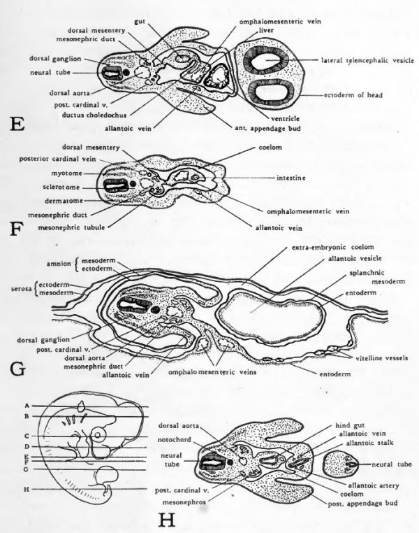
96 Hour Chick Embryo Serial Section
In the stage from 22 hours on, the somites formed in the mesoderm at the left and right side of the neural walls become visible. After 24 hours 4 to 5 segmented paired blocks can be discerned. Later on, these structures will differentiate into the vertebrae, the ribs, a part of the skin and the dorsal muscles.
48 hour chick serial sections. Lab practical. Thin layer of tissue surrounding the amnion. Exchanges gases and helps to provide oxygen to the embryo. In the chick this also brings calcium from the eggshell to the embryo in order to form the skeleton and beak. In mammals this forms part of the placenta. Derived from somatopleure. EE12-3 Chick Embryo 96 Hours Serial Sag Section Prepared Microscope Slide 96 hr chick; serial sagital section A 10% discount applies if you order more than 10 of this item and 15% discount applies if you order more than 25 of this item.
Only the head region lifts up above the area pellucida. In this preparation, one can see the chorda (notochord) in the region of the anterior intestinal portal. This structure marks the differentiating foregut which is formed as a blind pocket bordered by endodermal tissue. The neural walls end in a neural pore at the anterior side and become smaller and wider apart in the region of Hensen’s node where it ends in the sinus rhomboidalis. Sometimes the extra-embryonic vessels become already visible in the area vasculosa. Later on, they will make contact with the vitelline (omphalomesenteric) veins and arteries formed in the embryo. • Developmental stages after 22-28 hrs, according to Patten (1920) • Whole mount preparation 24 hours () • Cross sections 24 hours () Developemental stages 22-28 hrs according to Patten (1920) Dorsal view of a developing chicken embryo (between 22 - 28 hrs after fertilization) • 22 to 23 hrs: the beginning of somite formation • 24 hrs: 4 pairs of mesodermic somites are visible • 27-28 hrs: 8 pairs of mesodermic somites are visible Stage 24 hours Whole mount preparation 24 hours Information: The somites are formed in the mesoderm at the left and right side of the neural walls.
In this stage, they are visible as 4 to 5 segmented paired blocks. Afterwards these structures will differentiate in to the vertebrae, the ribs, a part of the skin and the dorsal muscles. Only this head region elevates above the underlying area pellucida. In this preparation, one can see the chorda (notochord) in the region of the differentiating foregut. Norfolk southern locomotive engineer training handbook manual. Embryology of the chicken 24 hours after fertilization Right: stained whole mount preparation. Herebelow A and B: cross sections at the level of the primitieve groove and the neural groove.
